Fichier:Frequency mapping in human ear and brain - 10.1371 journal.pbio.0030137.g001-L.jpg

Taille de cet aperçu : 488 × 599 pixels. Autres résolutions : 195 × 240 pixels | 391 × 480 pixels | 625 × 768 pixels | 834 × 1 024 pixels | 2 020 × 2 480 pixels.
Fichier d’origine (2 020 × 2 480 pixels, taille du fichier : 989 kio, type MIME : image/jpeg)
Ce fichier et sa description proviennent de Wikimedia Commons.

|
Une version vectorielle de cette image existe, dans le format « SVG ». Si elle n’est pas inférieure, elle devrait être utilisée à la place de la présente version pour des affichages en plus grandes dimensions ou nécessitant une meilleure résolution.
File:Frequency mapping in human ear and brain - 10.1371 journal.pbio.0030137.g001-L.jpg → File:Auditory Cortex Frequency Mapping.svg
Pour plus d’informations sur les images vectorielles, consultez la page de transition de Commons vers le format SVG. Voir aussi les informations à propos de la manière dont le logiciel MediaWiki gère les images au format SVG. |
 |
|
|
Une version vectorielle de cette image existe, dans le format « SVG ». Si elle n’est pas inférieure, elle devrait être utilisée à la place de la présente version pour des affichages en plus grandes dimensions ou nécessitant une meilleure résolution.
File:Frequency mapping in human ear and brain - 10.1371 journal.pbio.0030137.g001-L.jpg → File:Anatomy of Human Ear with Cochlear Frequency Mapping.svg
Pour plus d’informations sur les images vectorielles, consultez la page de transition de Commons vers le format SVG. Voir aussi les informations à propos de la manière dont le logiciel MediaWiki gère les images au format SVG. |
 |
Description
| Description |
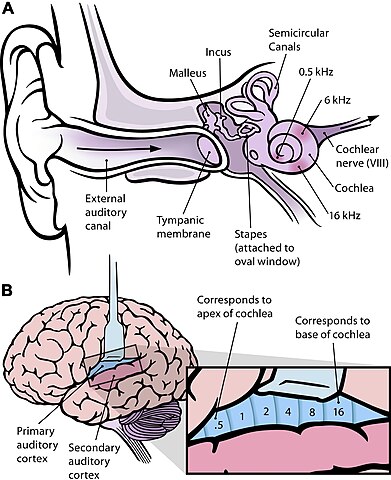
English: (A) The human ear and frequency mapping in the cochlea. The three ossicles incus, malleus, and stapes transmit airborne vibration from the tympanic membrane to the oval window at the base of the cochlea. Because of the mechanical properties of the basilar membrane within the snail-shaped cochlea, high frequencies will produce a vibration peak near the oval window, whereas low frequencies will stimulate receptors near the apex of the cochlea (locations for three frequencies indicated schematically). Information from the cochlear receptor cells is transmitted to the cochlear nuclei via the 8th cranial nerve, and on through the midbrain to the cortex. (Redrawn from Figure 12.3 in [11].)
(B) Lateral view of the human brain, with the auditory cortex exposed. The primary auditory cortex contains a topographic map of the cochlear frequency spectrum (shown in kilohertz). (Redrawn from Figure 12.15A in [11].) |
| Date | |
| Source | Perception Space—The Final Frontier, A PLoS Biology Vol. 3, No. 4, e137 doi:10.1371/journal.pbio.0030137 ([1]/[2]) |
| Auteur | Chittka L. and Brockmann A. |
| Autres versions |
Œuvres dérivées de ce fichier : |
![[2]](http://biology.plosjournals.org/archive/1545-7885/3/4/figure/10.1371_journal.pbio.0030137.g001-L.jpg)
Conditions d’utilisation
Ce fichier est disponible selon les termes de la licence Creative Commons Attribution 2.5 Générique.
- Vous êtes libre :
- de partager – de copier, distribuer et transmettre cette œuvre
- d’adapter – de modifier cette œuvre
- Sous les conditions suivantes :
- paternité – Vous devez donner les informations appropriées concernant l'auteur, fournir un lien vers la licence et indiquer si des modifications ont été faites. Vous pouvez faire cela par tout moyen raisonnable, mais en aucune façon suggérant que l’auteur vous soutient ou approuve l’utilisation que vous en faites.
Historique du fichier
Cliquer sur une date et heure pour voir le fichier tel qu'il était à ce moment-là.
| Date et heure | Vignette | Dimensions | Utilisateur | Commentaire | |
|---|---|---|---|---|---|
| actuel | 28 avril 2009 à 23:57 | | 2 020 × 2 480 (989 kio) | Mike.lifeguard | malleus and incus were swapped |
| 12 février 2009 à 06:06 |  | 2 020 × 2 480 (472 kio) | Mike.lifeguard | {{Information |Description={{en|1=(A) The human ear and frequency mapping in the cochlea. The three ossicles incus, malleus, and stapes transmit airborne vibration from the tympanic membrane to the oval window at the base of the cochlea. Because of the me |
Utilisation du fichier
Les 2 pages suivantes utilisent ce fichier :
Usage global du fichier
Les autres wikis suivants utilisent ce fichier :