Fichier:Coronavirus. SARS-CoV-2.png
Fichier d’origine(2 048 × 2 048 pixels, taille du fichier : 4,54 Mio, type MIME : image/png)

Ce fichier et sa description proviennent de Wikimedia Commons.

Description

Description
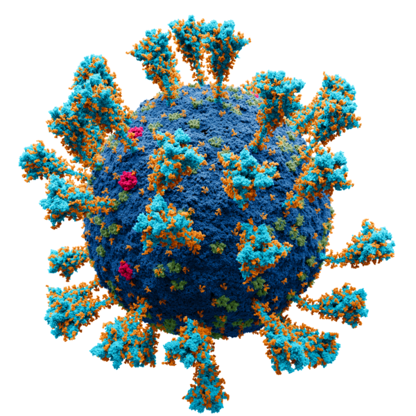
Deutsch: Wissenschaftlich genaues Atommodell der äußeren Struktur des SARS Coronavirus 2 (SARS-CoV-2), einem Stamm (genetische Variante) des Coronavirus, der die Coronavirus-Krankheit (COVID-19) verursachte und erstmals im Dezember 2019 in Wuhan, China, identifiziert wurde.

Jeder einzelne Ort (amorpher Fleck) ist ein Atom von:

 
kobalt: Virushülle
 
purpurrot: Hüllproteine
 
grün: Matrixproteine
 
orange: Glucose (Glycane)
 
türkis: Spike-Proteine
English: Scientifically accurate atomic model of the external structure of the Severe Acute Respiratory Syndrome CoronaVirus 2 (SARS-CoV-2), a strain (genetic variant) of the coronavirus that caused coronavirus disease (COVID-19), first identified in Wuhan, China, during December 2019

Each separate locus (amorphous blob) is a molecule of:

 
cobalt: membrane
 
crimson: E protein
 
green: M protein
 
orange: glucose (glycan)
 
turquoise : S (spike) glycoprotein
Español: Modelo atómico de la estructura externa del SARS-CoV-2. Cada "bola" es un átomo.
Leyenda:
 
azul cobalto — membrana
 
turquesa — glicoproteína de pico (S)
 
carmesí — proteína E
 
verde — proteína M
 
naranja — glucosa
Русский: Научно достоверная атомарная модель внешней структуры коронавируса (SARS-CoV-2). Каждый "шарик" — атом. Опубликовано на N+1.

Научный консультант:
Никитин Н. А. (доктор биологических наук, специалист в области вирусологии)
Борисевич С.С. к.х.н специалист по молекулярному моделированию поверхностных вирусных белков, руководитель группы «Кванты и Динамика», (с.н.с. лаборатория химической физики УфИХ РАН)
Архипова В.И. (специалитет по направлению фундаментальная и прикладная химия. Лаборатория химии РНК, Институт химической биологии и фундаментальной медицины Сибирского отделения Российской академии наук).

  • В работе были использованы следующие структуры из открытых источников:

Protein Data Bank: 2mls, 6y3y, 5x29, 6yyt Charmm-gui: 6vsb_1_1_1_S309

  • Цветовое обозначение:
 
кобальт — мембрана.
 
бирюза — S-белок.
 
малиновый — E-белок.
 
зелёный — M-белок.
 
оранжевый — гликаны.
Проект был сделан в соответствии с научными источниками:
  1. Surya, W., Li, Y., Torres, J. Structural model of the SARS coronavirus E channel in LMPG micelles. // Biochim. Biophys. Acta - Biomembr. – 2018. – Vol. 1860. – N. 6. – P. 1309–1317.
  2. Koppisetti, R. K., Fulcher, Y. G., Jurkevich, A., Prior, S. H., Xu, J., Lenoir, M., Overduin, M., Van Doren, S. R. Ambidextrous binding of cell and membrane bilayers by soluble matrix metalloproteinase-12. // Nat. Commun. – 2014. – Vol. 5. – P. 1–14.
  3. Hillen, H. S., Kokic, G., Farnung, L., Dienemann, C., Tegunov, D., Cramer, P. Structure of replicating SARS-CoV-2 polymerase. // Nature. – 2020. – Vol. 584. – N. 7819. – P. 154–156.
  4. Harris, L. J., Larson, S. B., Hasel, K. W., McPherson, A. Refined structure of an intact IgG2a monoclonal antibody. // Biochemistry. – 1997. – Vol. 36. – N. 7. – P. 1581–1597.
  5. Noreng, S., Bharadwaj, A., Posert, R., Yoshioka, C., Baconguis, I. Structure of the human epithelial sodium channel by cryo-electron microscopy. // Elife. – 2018. – Vol. 7. – P. 1–23.
  6. Almond, A., DeAngelis, P. L., Blundell, C. D. Hyaluronan: The Local Solution Conformation Determined by NMR and Computer Modeling is Close to a Contracted Left-handed 4-Fold Helix. // J. Mol. Biol. – 2006. – Vol. 358. – N. 5. – P. 1256–1269.
  7. Hurdiss, D. L., Drulyte, I., Lang, Y., Shamorkina, T. M., Pronker, M. F., van Kuppeveld, F. J. M., Snijder, J., de Groot, R. J. Cryo-EM structure of coronavirus-HKU1 haemagglutinin esterase reveals architectural changes arising from prolonged circulation in humans. // Nat. Commun. – 2020. – Vol. 11. – N. 1. – P. 1–10.
  8. Yan, Renhong, Yuanyuan Zhang, Yaning Li, Lu Xia, Yingying Guo, Q. Z. Structural basis for the recognition of SARS-CoV-2 by full-length human ACE2. // Science (80-. ). – 2020. – Vol. 3. – N. 3. – P. 1–8.
  9. Javitt, G., Khmelnitsky, L., Albert, L., Bigman, L. S., Elad, N., Morgenstern, D., Ilani, T., Levy, Y., Diskin, R., Fass, D. Assembly Mechanism of Mucin and von Willebrand Factor Polymers. // Cell. – 2020. – Vol. 183. – N. 3. – P. 717-729.e16.
  10. Daniel Wrapp, Nianshuang Wang, Kizzmekia S. Corbett, Jory A. Goldsmith, Ching-Lin Hsieh, Olubukola Abiona, B. S. G., McLellan, and J. S. Cryo-EM structure of the 2019-nCoV spike in the prefusion conformation. // Science (80-. ). – 2020. – Vol. 21. – N. 1. – P. 1–9.
  11. Wang, M. Y., Zhao, R., Gao, L. J., Gao, X. F., Wang, D. P., Cao, J. M. SARS-CoV-2: Structure, Biology, and Structure-Based Therapeutics Development. // Front. Cell. Infect. Microbiol. – 2020. – Vol. 10. – N. November. – P. 1–17.
  12. Yao, H., Song, Y., Chen, Y., Wu, N., Xu, J., Sun, C., Zhang, J., Weng, T., Zhang, Z., Wu, Z., Cheng, L., Shi, D., Lu, X., Lei, J., Crispin, M., Shi, Y., Li, L., Li, S. Molecular Architecture of the SARS-CoV-2 Virus. // Cell. – 2020. – Vol. 183. – N. 3. – P. 730-738.e13.
  13. Oostra, M., de Haan, C. A. M., de Groot, R. J., Rottier, P. J. M. Glycosylation of the Severe Acute Respiratory Syndrome Coronavirus Triple-Spanning Membrane Proteins 3a and M. // J. Virol. – 2006. – Vol. 80. – N. 5. – P. 2326–2336.
  14. B.W. Neuman, M. J. B. Supramolecular Architecture of the Coronavirus Particle. // Adv. Virus Res. – 2020. – Vol. 96. – P. 1–27.
  15. Neuman, B. W., Kiss, G., Kunding, A. H., Bhella, D., Baksh, M. F., Connelly, S., Droese, B., Klaus, J. P., Makino, S., Sawicki, S. G., Siddell, S. G., Stamou, D. G., Wilson, I. A., Kuhn, P., Buchmeier, M. J. A structural analysis of M protein in coronavirus assembly and morphology. // J. Struct. Biol. – 2011. – Vol. 174. – N. 1. – P. 11–22.
  16. Yu, A., Pak, A. J., He, P., Monje-Galvan, V., Casalino, L., Gaieb, Z., Dommer, A. C., Amaro, R. E., Voth, G. A. A multiscale coarse-grained model of the SARS-CoV-2 virion. // Biophys. J. – 2021. – Vol. 120. – N. 6. – P. 1097–1104.
  17. Yao, H., Song, Y., Chen, Y., Wu, N., Xu, J., Sun, C., Zhang, J., Weng, T., Zhang, Z., Wu, Z., Cheng, L., Shi, D., Lu, X., Lei, J., Crispin, M., Shi, Y., Li, L., Li, S. Molecular architecture of the SARS-CoV-2 virus. // Cell. – 2020. – Vol. 183. – N. 3. – P. 730–738.
  18. Choi, Y. K., Cao, Y., Frank, M., Woo, H., Park, S. J., Yeom, M. S., Croll, T. I., Seok, C., Im, W. Structure, Dynamics, Receptor Binding, and Antibody Binding of the Fully Glycosylated Full-Length SARS-CoV-2 Spike Protein in a Viral Membrane. // J. Chem. Theory Comput. – 2021. – Vol. 17. – N. 4. – P. 2479–2487.

Первичные источники:


Производные моделей протеинов созданы на базе моделей со свободной лицензией (freely available for both non-commercial and commercial use).
Українська: Атомарна модель зовнішньої структури коронавірусу SARS-CoV-2. Кожен «кулька» — атом.
Date
Source

Travail personnel. Scientific consultants:

Nikitin N.A., Doctor of Biological Sciences, Department of Virology, Faculty of Biology, Lomonosov Moscow State University.
Borisevich S.S. Candidate of Chemical Sciences, Specialist in Molecular Modeling of Viral Surface Proteins, Senior Researcher, Head of the "Quantum & Dynamics»,Laboratory of Chemical Physics, Ufa Institute of Chemistry RAS
Arkhipova V.I., specialization in Fundamental and Applied chemistry, senior engineer, RNA Chemistry Laboratory, Institute of chemical biology and fundamental medicine SB RAS
Auteur Alexey Solodovnikov (Idea, Producer, CG, Editor), Valeria Arkhipova (Scientific Сonsultant)
Autorisation
(Réutilisation de ce fichier)

Published by N+1 a popular science online publication of Russia (https://nplus1.ru/), protein models are derivative works of the

free license site (freely available for both non-commercial and commercial use)
Autres versions

Sources

Primary sources:

The following structures from open sources were used in the work, Protein Data Bank (https://www.rcsb.org):

Additional sources:

  1. Surya, W., Li, Y., Torres, J. Structural model of the SARS coronavirus E channel in LMPG micelles // Biochim. Biophys. Acta - Biomembr. – 2018. – Vol. 1860. – N. 6. – P. 1309–1317.
  2. Koppisetti, R. K., Fulcher, Y. G., Jurkevich, A., Prior, S. H., Xu, J., Lenoir, M., Overduin, M., Van Doren, S. R. Ambidextrous binding of cell and membrane bilayers by soluble matrix metalloproteinase-12 // Nat. Commun. – 2014. – Vol. 5. – P. 1–14.
  3. Hillen, H. S., Kokic, G., Farnung, L., Dienemann, C., Tegunov, D., Cramer, P. Structure of replicating SARS-CoV-2 polymerase // Nature. – 2020. – Vol. 584. – N. 7819. – P. 154–156.
  4. Harris, L. J., Larson, S. B., Hasel, K. W., McPherson, A. Refined structure of an intact IgG2a monoclonal antibody // Biochemistry. – 1997. – Vol. 36. – N. 7. – P. 1581–1597.
  5. Noreng, S., Bharadwaj, A., Posert, R., Yoshioka, C., Baconguis, I. Structure of the human epithelial sodium channel by cryo-electron microscopy // Elife. – 2018. – Vol. 7. – P. 1–23.
  6. Almond, A., DeAngelis, P. L., Blundell, C. D. Hyaluronan: The Local Solution Conformation Determined by NMR and Computer Modeling is Close to a Contracted Left-handed 4-Fold Helix // J. Mol. Biol. – 2006. – Vol. 358. – N. 5. – P. 1256–1269.
  7. Hurdiss, D. L., Drulyte, I., Lang, Y., Shamorkina, T. M., Pronker, M. F., van Kuppeveld, F. J. M., Snijder, J., de Groot, R. J. Cryo-EM structure of coronavirus-HKU1 haemagglutinin esterase reveals architectural changes arising from prolonged circulation in humans // Nat. Commun. – 2020. – Vol. 11. – N. 1. – P. 1–10.
  8. Yan, Renhong, Yuanyuan Zhang, Yaning Li, Lu Xia, Yingying Guo, Q. Z. Structural basis for the recognition of SARS-CoV-2 by full-length human ACE2 // Science (80-. ). – 2020. – Vol. 3. – N. 3. – P. 1–8.
  9. Javitt, G., Khmelnitsky, L., Albert, L., Bigman, L. S., Elad, N., Morgenstern, D., Ilani, T., Levy, Y., Diskin, R., Fass, D. Assembly Mechanism of Mucin and von Willebrand Factor Polymers // Cell. – 2020. – Vol. 183. – N. 3. – P. 717-729.e16.
  10. Daniel Wrapp, Nianshuang Wang, Kizzmekia S. Corbett, Jory A. Goldsmith, Ching-Lin Hsieh, Olubukola Abiona, B. S. G., McLellan, and J. S. Cryo-EM structure of the 2019-nCoV spike in the prefusion conformation // Science (80-. ). – 2020. – Vol. 21. – N. 1. – P. 1–9.
  11. Wang, M. Y., Zhao, R., Gao, L. J., Gao, X. F., Wang, D. P., Cao, J. M. SARS-CoV-2: Structure, Biology, and Structure-Based Therapeutics Development // Front. Cell. Infect. Microbiol. – 2020. – Vol. 10. – N. November. – P. 1–17. (https://pubmed.ncbi.nlm.nih.gov/33324574/)
  12. Yao, H., Song, Y., Chen, Y., Wu, N., Xu, J., Sun, C., Zhang, J., Weng, T., Zhang, Z., Wu, Z., Cheng, L., Shi, D., Lu, X., Lei, J., Crispin, M., Shi, Y., Li, L., Li, S. Molecular Architecture of the SARS-CoV-2 Virus // Cell. – 2020. – Vol. 183. – N. 3. – P. 730-738.e13.
  13. Oostra, M., de Haan, C. A. M., de Groot, R. J., Rottier, P. J. M. Glycosylation of the Severe Acute Respiratory Syndrome Coronavirus Triple-Spanning Membrane Proteins 3a and M // J. Virol. – 2006. – Vol. 80. – N. 5. – P. 2326–2336. (https://europepmc.org/article/MED/16474139)
  14. B.W. Neuman, M. J. B. Supramolecular Architecture of the Coronavirus Particle // Adv. Virus Res. – 2020. – Vol. 96. – P. 1–27 (https://www.ncbi.nlm.nih.gov/pmc/articles/PMC7112365/, https://europepmc.org/article/PMC/1563832)
  15. Neuman, B. W., Kiss, G., Kunding, A. H., Bhella, D., Baksh, M. F., Connelly, S., Droese, B., Klaus, J. P., Makino, S., Sawicki, S. G., Siddell, S. G., Stamou, D. G., Wilson, I. A., Kuhn, P., Buchmeier, M. J. A structural analysis of M protein in coronavirus assembly and morphology // J. Struct. Biol. – 2011. – Vol. 174. – N. 1. – P. 11–22. (https://www.ncbi.nlm.nih.gov/pmc/articles/PMC4486061/)
  16. Yu, A., Pak, A. J., He, P., Monje-Galvan, V., Casalino, L., Gaieb, Z., Dommer, A. C., Amaro, R. E., Voth, G. A. A multiscale coarse-grained model of the SARS-CoV-2 virion // Biophys. J. – 2021. – Vol. 120. – N. 6. – P. 1097–1104 (https://europepmc.org/article/PMC/PMC7695975, https://search.bvsalud.org/global-literature-on-novel-coronavirus-2019-ncov/resource/en/covidwho-947143)
  17. Yao, H., Song, Y., Chen, Y., Wu, N., Xu, J., Sun, C., Zhang, J., Weng, T., Zhang, Z., Wu, Z., Cheng, L., Shi, D., Lu, X., Lei, J., Crispin, M., Shi, Y., Li, L., Li, S. Molecular architecture of the SARS-CoV-2 virus // Cell. – 2020. – Vol. 183. – N. 3. – P. 730–738 (https://www.sciencedirect.com/science/article/pii/S0092867420311594)
  18. Choi, Y. K., Cao, Y., Frank, M., Woo, H., Park, S. J., Yeom, M. S., Croll, T. I., Seok, C., Im, W. Structure, Dynamics, Receptor Binding, and Antibody Binding of the Fully Glycosylated Full-Length SARS-CoV-2 Spike Protein in a Viral Membrane // J. Chem. Theory Comput. – 2021. – Vol. 17. – N. 4. – P. 2479–2487 (https://www.researchgate.net/publication/349986293_Structure_Dynamics_Receptor_Binding_and_Antibody_Binding_of_the_Fully_Glycosylated_Full-Length_SARS-CoV-2_Spike_Protein_in_a_Viral_Membrane)

Conditions d’utilisation

Moi, en tant que détenteur des droits d’auteur sur cette œuvre, je la publie sous la licence suivante :
w:fr:Creative Commons
paternité partage à l’identique
Vous êtes libre :
  • de partager – de copier, distribuer et transmettre cette œuvre
  • d’adapter – de modifier cette œuvre
Sous les conditions suivantes :
  • paternité – Vous devez donner les informations appropriées concernant l'auteur, fournir un lien vers la licence et indiquer si des modifications ont été faites. Vous pouvez faire cela par tout moyen raisonnable, mais en aucune façon suggérant que l’auteur vous soutient ou approuve l’utilisation que vous en faites.
  • partage à l’identique – Si vous modifiez, transformez, ou vous basez sur cette œuvre, vous devez distribuer votre contribution sous la même licence ou une licence compatible avec celle de l’original.

Distinctions

Image de l'année
Image de l'année
Image du jour
Image du jour
Image remarquable
Image de qualité
Image de qualité
Image de valeur
Image de valeur

Wikimedia Commons

Ce fichier a été proposé en tant que finaliste de l’image de l’année 2021.
Ce fichier a été image du jour le 8 août 2021.
Ceci est une image remarquable sur Wikimedia Commons (Images remarquables) et est considéré(e) comme l'une de nos meilleures images.
Cette image est une image de qualité et est considérée comme respectant le guide des images.
Ceci est une image de valeur et est considérée comme remplissant les critères des images de valeur.

Si vous avez une image de qualité similaire qui peut être publiée sous une licence compatible, vous pouvez l’importer, y apposer le bandeau de licence adéquat, et la proposer.

Légendes

Modèle atomique scientifiquement exact du coronavirus (SARS-CoV-2). Chaque "balle" représente un atome.

Éléments décrits dans ce fichier

dépeint

image/png

eb4db5448ad22fc4c229437fae4692de527c749d

4 765 767 octet

2 048 pixel

2 048 pixel

Historique du fichier

Cliquer sur une date et heure pour voir le fichier tel qu'il était à ce moment-là.

Date et heureVignetteDimensionsUtilisateurCommentaire
actuel10 janvier 2022 à 00:17Vignette pour la version du 10 janvier 2022 à 00:172 048 × 2 048 (4,54 Mio)Jul059Lossless file size reduction
24 septembre 2021 à 05:58Vignette pour la version du 24 septembre 2021 à 05:582 048 × 2 048 (4,6 Mio)Iketsilossless compression
15 juin 2021 à 18:06Vignette pour la version du 15 juin 2021 à 18:062 048 × 2 048 (5,34 Mio)AlexeySolodovnikovfix color bug
13 juin 2021 à 16:28Vignette pour la version du 13 juin 2021 à 16:282 048 × 2 048 (5,34 Mio)AlexeySolodovnikovМы обновили модель. В роли нашего научного консультанта выступил доктор биологических наук, специалист в области вирусологии, Никитин Н. А. и к.х.н специалист по молекулярному моделированию поверхностных вирусных белков Борисевич С.С. Под их руководством в модель были внесены следующие правки: Изменено количество S-белков с 90 до 38, количество M-белков было увеличено до 1000, а E-белков, как минорных компонентов мембраны, снижено до 15, HE-белок удалён. Также была принята во внимание шарни...
17 mai 2021 à 13:06Vignette pour la version du 17 mai 2021 à 13:062 048 × 2 048 (16,04 Mio)AlexeySolodovnikovadd alpha
4 mai 2021 à 20:41Vignette pour la version du 4 mai 2021 à 20:412 048 × 2 048 (16,04 Mio)AlexeySolodovnikovUploaded own work with UploadWizard

Usage global du fichier

Les autres wikis suivants utilisent ce fichier :

Voir davantage sur l’utilisation globale de ce fichier.