Fichier:Blood clot in scanning electron microscopy.jpg
Pas de plus haute résolution disponible.
Blood_clot_in_scanning_electron_microscopy.jpg (700 × 475 pixels, taille du fichier : 76 kio, type MIME : image/jpeg)
Ce fichier et sa description proviennent de Wikimedia Commons.
Description
| Description |
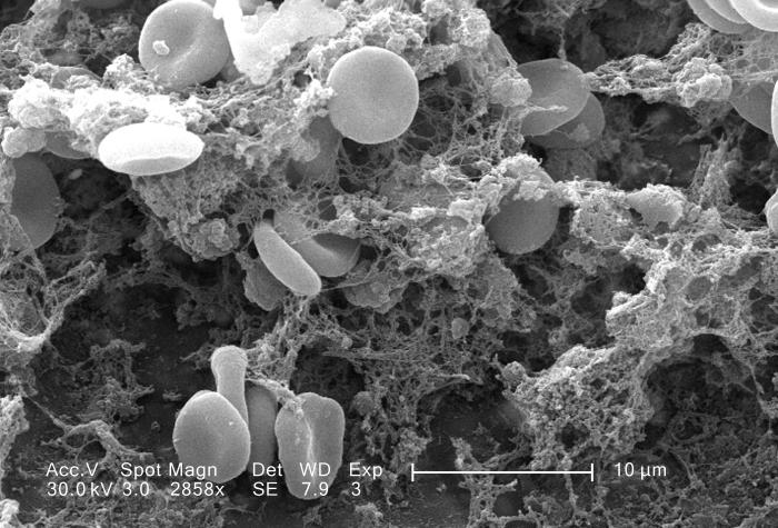
English: This scanning electron micrograph (SEM) depicted a number of red blood cells found enmeshed in a fibrinous matrix on the luminal surface of an indwelling vascular catheter; Magnified 2858x.
Note the biconcave cytomorphologic shape of each erythrocyte, which increases the surface area of these hemoglobin-filled cells, thereby, promoting a greater degree of gas exchange, which is their primary function in an in vivo setting. In their adult phase, these cells possess no nucleus. What appears to be irregularly-shaped chunks of debris, are actually fibrin clumps, which when inside the living organism, functions as a key component in the process of blood clot formation, acting to entrap the red blood cells in a mesh-like latticework of proteinaceous strands, thereby, stabilizing and strengthening the clot, in much the same way as rebar acts to strengthen, and reinforce cement. |
| Date | |
| Source | http://phil.cdc.gov/phil/details.asp |
| Auteur | Janice Carr |
Conditions d’utilisation
| Cette œuvre a été placée dans le domaine public par son auteur, http://phil.cdc.gov/phil/details.asp Janice Carr. Ceci s’applique dans le monde entier. Dans certains pays, ceci peut ne pas être possible ; dans ce cas : http://phil.cdc.gov/phil/details.asp Janice Carr accorde à toute personne le droit d’utiliser cette œuvre dans n’importe quel but, sans aucune condition, sauf celles requises par la loi.
|
Historique du fichier
Cliquer sur une date et heure pour voir le fichier tel qu'il était à ce moment-là.
| Date et heure | Vignette | Dimensions | Utilisateur | Commentaire | |
|---|---|---|---|---|---|
| actuel | 13 mai 2015 à 19:25 | | 700 × 475 (76 kio) | Jean-madeleine de sainte agathe | User created page with UploadWizard |
Utilisation du fichier
La page suivante utilise ce fichier :
Usage global du fichier
Les autres wikis suivants utilisent ce fichier :