Fichier:Annals of tropical medicine and parasitology (1907) (14799033113).jpg

Fichier d’origine(2 022 × 3 194 pixels, taille du fichier : 1,35 Mio, type MIME : image/jpeg)

Ce fichier et sa description proviennent de Wikimedia Commons.

Description

Description
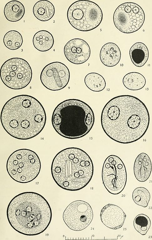
English:

Identifier: annalsoftropical12live (find matches)
Title: Annals of tropical medicine and parasitology
Year: 1907 (1900s)
Authors: Liverpool School of Tropical Medicine
Subjects: Parasites
Publisher: Liverpool University Press (etc.)
Contributing Library: Gerstein - University of Toronto
Digitizing Sponsor: University of Toronto

View Book Page: Book Viewer
About This Book: Catalog Entry
View All Images: All Images From Book
Click here to view book online to see this illustration in context in a browseable online version of this book.

Text Appearing Before Image:

EXPLANATION OF PLATE

   All the figures (except 9 and 24) were drawn, from cysts stained With iodine, with the aid of a camera lucida at a magnification of 3,300. T hey have been reduced in reproduction.
Figs. 1-9. Cysts of Entamoeba histolytica.

Fig. 1. Uninucleate cyst containing vacuole. Diameter 8 µ
Fig. 2. Uninucleate cyst with a single chromatoid body. Diameter 8 µ
Fig. 3. Binucleate cyst. Diameter 7.5 µ
Fig. 4. Tetranucleate cyst. Diameter 8 µ
Fig. 5. Uninucleate cyst with diffuse staining vacuole, size 13.5 µ × 12 µ
Fig. 6. Binucleate cyst with diffuse staining vacuole. Diameter 12 µ
Fig. 7. Binucleate cyst with several vacuoles unstained by iodine. Diameter 10 µ
Fig. 8. Tetranucleate. cyst. Diameter 12.5 µ
Fig. 9. Binucleate cyst With typical chromatoid body. Diameter 11 µ. Cyst as seen in saline.
Figs. 10-13. Cysts of Entamoeba nana.
(rem.: this species has been re-classified as Endolimax nana now).
Fig. 10. Tetranucleate cyst, nuclei remarkably clear. Size 9 µ × 8 µ
Fig. 11. Binucleate cyst with deeply stained mass of glycogen. Size 9 µ × 7.5 µ
Fig. 12. Binucleate cyst, nuclei faintly stained. Size 8.5 µ × 7 µ
Fig. 13. Uninucleate cyst, nucleus faintly stained. Size 8.5 µ × 7 µ
In all four cysts the refringent granules of volutin were clearly discernible.
Figs. 14-19. Cysts of Entamoeba coli.
Fig. 14. Tetranucleate cyst. Diameter 17.5 µ
Fig. 15. Binucleate cyst with large deeply stained centrally placed vacuole. Diameter 16 µ
Fig. 16. Binucleate cyst without vacuole. Diameter 18.5 µ
Fig. 17. Eight nucleate cyst. Diameter 13 µ
Fig. 18. Eight nucleate cyst with pointed chromatoid bodies. Diameter 16 µ
Fig. 19. Probably abnormal eight nucleate cyst. Diameter 17 µ
Figs. 20-21. Cysts of Giardia intestinalis.
Fig. 20. Large brown staining cyst. Size 15 µ × 9.5nbsp;µ
Fig. 21. Small bluish-grey staining cyst. Size lO µ × 6.5/1
Figs. 22-23. Cysts of Chilomastix mesnili.
Fig. 22. The cytostome and nucleus clearly visible. Size 8.5 µ × 7.5 µ
Fig. 23. The cytostome and nucleus partially obscured by large deeply stainingmass of glycogen. Size 8.5 µ × 7.5 µ
Fig. 24. Iodine cyst drawn from specimen stained with iron haematoxylin. The clear space would appear dark brown in colour if stained with iodine and the nucleus would be less sharply defined. Diameter 11.5 µ
Fig. 25. Blastocystis hominis. Diameter 10 µ


Annals Trop. Med. & Parasitol., Vol. XII              PLATE I


Text Appearing After Image:
J. R. M., del. C. Tinlins & Co., Ltd., Imp. 27 MEASUREMENTS OF AND OBSERVA-TIONS UPON THE CYSTS OFENTAMOEBA HISTOLYTICA ANDOF ENTAMOEBA COLI BY A. MALINS SMITH, M.A. (Cantab.) From the Liverpool School of Tropical Medicine (^Received for publication 26 March, 1918) From April 22nd, 1917, until March 4th, 1918, the writermeasured the dimensions of 2,833 cysts of two of the intestinalentamoebae of man, E. histolytica and E. coli. The results of thesemeasurements are given in the present paper. At the time eachmeasurement was made the nuclei of the cyst were counted andtheir number recorded. A note was also made of the presence orabsence of chromatoid bodies and vacuoles. The main results ofthese observations are recorded here also. METHODS The majority of the stools from which the cysts to be measuredwere taken were those of convalescent dysenteries, and were sent tothe laboratory for the usual routine examination for dysentery.Cysts were also measured from a small number of carrier

Note About Images

Please note that these images are extracted from scanned page images that may have been digitally enhanced for readability - coloration and appearance of these illustrations may not perfectly resemble the original work.
Date
Source

https://www.flickr.com/photos/internetarchivebookimages/14799033113/

Auteur Liverpool School of Tropical Medicine
Autorisation
(Réutilisation de ce fichier)
At the time of upload, the image license was automatically confirmed using the Flickr API. For more information see Flickr API detail.
Autres versions
Volume
InfoField
12
Flickr tags
InfoField
  • bookid:annalsoftropical12live
  • bookyear:1907
  • bookdecade:1900
  • bookcentury:1900
  • bookauthor:Liverpool_School_of_Tropical_Medicine
  • booksubject:Parasites
  • bookpublisher:Liverpool_University_Press__etc__
  • bookcontributor:Gerstein___University_of_Toronto
  • booksponsor:University_of_Toronto
  • bookleafnumber:42
  • bookcollection:gerstein
  • bookcollection:toronto
  • bookcollection:medicalheritagelibrary
  • BHL Collection
Flickr posted date
InfoField
29 juillet 2014


Conditions d’utilisation

Cette image est issue de la collection The Commons du site Flickr. Les organismes y partageant leur collection déclarent qu'à leur connaissance, aucune restriction de droit d'auteur ne fait obstacle à leur diffusion, pour l'une des raisons suivantes :
  1. l'œuvre est dans le domaine public car les droits d'auteur ont expiré ;
  2. l'œuvre a été rattachée au domaine public pour d'autres raisons, tel que le non respect des conditions nécessaires pour renouveler des droits d'auteur ;
  3. l'organisme est détenteur des droits d'auteur mais ne ne souhaite pas exercer son contrôle ou ;
  4. l'organisme possède des droits suffisant pour permettre à des tiers d'utiliser ces travaux sans aucune restriction.

La page https://flickr.com/commons/usage/ donne à ce sujet plus d'informations.


Merci d'ajouter des bandeaux de licence supplémentaires à cette image si des informations plus spécifiques sont disponibles à propos du statut de cette image. Consultez Commons:À propos des licences pour plus d'informations.
Cette image a été originellement postée sur Flickr par Internet Archive Book Images à l'adresse https://flickr.com/photos/126377022@N07/14799033113. Elle a été passée en revue le 15 septembre 2015 par le robot FlickreviewR, qui a confirmé qu'elle se trouvait sous licence No known copyright restrictions.

15 septembre 2015

Légendes

Ajoutez en une ligne la description de ce que représente ce fichier

Éléments décrits dans ce fichier

dépeint

Valeur sans élément de Wikidata

auteur ou autrice (chaîne) : Liverpool School of Tropical Medicine

image/jpeg

e5c411618cdcec27afe2e159554cf389bf0cb729

1 413 459 octet

3 194 pixel

2 022 pixel

Historique du fichier

Cliquer sur une date et heure pour voir le fichier tel qu'il était à ce moment-là.

Date et heureVignetteDimensionsUtilisateurCommentaire
actuel15 septembre 2015 à 10:53Vignette pour la version du 15 septembre 2015 à 10:532 022 × 3 194 (1,35 Mio)== {{int:filedesc}} == {{information |description={{en|1=<br> '''Identifier''': annalsoftropical12live ([https://commons.wikimedia.org/w/index.php?title=Special%3ASearch&profile=default&fulltext=Search&search=insource%3A%2Fannalsoftropical12live%2F fin...

Aucune page n’utilise ce fichier.